D - ESP
<< >>
Coronavirus19 5i: Dec.25, 2020: Dr. Vanessa Schmidt Video 2: Warning of lipid nanoparticles in genetic vaccinations (video 2)
Lipid nanoparticles destroy cell walls - cell death - thrombosis - multiple organ failure - ev. there is PEG prior charge provoking allergic shocks etc.
Video (December 25, 2020): What are the dangers of lipid nanoparticles? BioNTech vaccine (52min.)
(orig. German: Welche Gefahren gehen von den Lipid-Nanopartikeln aus ? BioNTech-Impfstoff)
Video link: https://www.youtube.com/watch?v=oNGFXiBVV8M
YouTube channel: Das Leben der Moleküle - Gesundheit verstehen ! - uploaded on Dec.25, 2020
What do you call the Corona19 policy? - OVERPROTECTION - Governments fuck off to psychiatry! (Michael Palomino, January 31, 2021)
How do you survive a vaccination? Just NEVER VACCINATE! (Michael Palomino, February 21, 2021)
resented by Michael Palomino (2021)
| Teilen / share: |
Facebook |
|
Twitter
|
|
|
|
Lockdown is COERCION + COMMERCIAL DAMAGE!
Mask terror is a COERCION + HEALTH HAZARD!
Refusal to provide information about natural medicine is FAILURE TO PROVIDE HELP!
The immune system can be supported with citrus fruits (Vit. C), with garlic + onions + ginger (natural antibiotics) + all food must be whole grain food (for minerals), as well as olive oil + nuts must be.
French fries (frit oil contaminated) and white rice + white flour (without minerals) and lemonade with citric acid (corrosive) have not yet been banned, this is really negligent.
Michael Palomino, March 18, 2020
-- The best prevention or cure for the flu is a medical diet + blood group nutrition + going to bed early + baking soda with apple cider vinegar (note sodium bicarbonate)
-- Previous illnesses usually heal away with sodium bicarbonate (note sodium bicarbonate).
May 17, 2020: Mossad government Merkel is DIRECTLY CONTROLLED by Bill Gates and Melinda Gates - through a "declaration of intent" from Feb. 2017 - Link
May 17, 2020: Mossad government in Switzerland is DIRECTLY CONTROLLED by Bill Gates+Melinda Gates - with a "declaration of intent", January 22, 2014 - Link
Uncut-News March 10, 2021: What dangers do lipid nanoparticles pose? BioNTech vaccine
(orig. German: Welche Gefahren gehen von den Lipid-Nanopartikeln aus ? BioNTech-Impfstoff
The article What dangers do lipid nanoparticles pose? BioNTech vaccine appeared on uncut-news.ch ...
https://uncutnews.ch/welche-gefahren-gehen-von-den-lipid-nanopartikeln-aus-biontech-impfstoff/
Dr. Vanessa Schmidt (cell microbiologist) during her speech about lipid nanoparticles [0]
Video (December 25, 2020): What are the dangers of lipid nanoparticles? BioNTech vaccine (52min.)
(orig. German: Welche Gefahren gehen von den Lipid-Nanopartikeln aus ? BioNTech-Impfstoff)
Video (December 25, 2020): What are the dangers of lipid nanoparticles? BioNTech vaccine (52min.)
(orig. German: Welche Gefahren gehen von den Lipid-Nanopartikeln aus ? BioNTech-Impfstoff)
https://www.youtube.com/watch?v=oNGFXiBVV8M - YouTube channel: Das Leben der Moleküle - Gesundheit verstehen ! - uploaded on Dec.25, 2020
Dr. Vanessa Schmidt-Krüger explains how the lipids in the body are deadly - and the deadly effect of polyethylene glycol (PEG):
from: https://www.youtube.com/watch?v=oNGFXiBVV8M
-- Minute 2 to 15: Components of the lipid nanoparticle (LNP) from Pfizer/BioNTech and functions of the individual components
-- Minute 15 to 20: Where in the body do the lipid nanoparticles (LNP) go?
-- Minute 20 to 35: The side effects of the Pfizer/BioNTech vaccine tried on rats - damage to the liver and at the muscle!
-- Minute 35 to 41: damage to the adaptive immune system.
-- Minute 41 to 48: Why do the body cells die from lipid nanoparticles (LNPs)?
-- Minute 48 to 50: Polyethylene glycol (PEG) and allergy
Summary: After the vesicle with mRNA bursts, lipids swim around freely + cause major to fatal problems after approx. 6 months - liver is destroyed etc.
Toxic Pfizer / BionTech injections are called 1) BNT162b1 - and a second version 2) BNT162b2
Around the mRNA, a protection envelope is installed with the lipids called ALC-0315 and ALC-0159 so that this mRNA is not broken down in the body. This unit ic called vesicle. This vesicle is manipulated to the cells are tearing it into the cells. In the cells, this mRNA vesicle is filled with water and then the vesicle bursts, the mRNA and the lipids swim around freely - and if you don't die of shock or the euthanasia agent potassium chloride, you will have severe to fatal problems in 6 months because these lipids then cause the most serious to fatal problems, including destroying the liver, etc. All of this has been proven in rat experiments when pharma industries wanted to develop a SARS vaccine since 2002 with rats, whereby these rat experiments are HIDDEN in the mainstream media.
And there is the PEG risk: If there is a hyperallergy to polyethylene glycol 2000 (PEG), the emergency unit is the next station of life immediately - possibly death.
The presentation (translation):
What is in there is absolutely the consequence of my fears (1'46 ''), which I already told you in the first video (1'51 ''), that is dangerous and, but you can decide for yourself (1'56''). Let's start (1'58 ''):
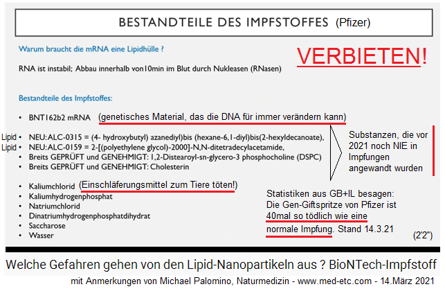
SLIDE 1: RNA needs lipid envelope, otherwise the RNA is "degraded" within 10 minutes - there are 4 lipids
(orig. German: FOLIE 1: RNA braucht Lipidhülle, sonst wird die RNA in 10 Minuten "abgebaut" - 4 Lipide)
Title: INGREDIENTS OF THE VACCINE (Pfizer)
Why does mRNA need a lipid envelope: RNA is unstable; Degradation in the blood within 10 minutes by nucleases (RNases). Components of the vaccine: BNT162b2 mRNA
-- [Lipid!]: NEW: ALC-0315 = (4-hydroxybutyl) azanediyl) bis (hexane-6, 1-diyl) bis (2-hexyldecanoate)
-- [Lipid!]: NEW: ALC-0159 = 2 - [(polyethylene glycol) -2000] -N, N-ditetradecylacetamide
-- Already TESTED and APPROVED: 1,2-Distearoyl-sn-glycero-3 phosphocholine (DSPC)
-- Already TESTED and APPROVED:
-- cholesterol
-- potassium chloride (Kaliumchlorid) [this is the killing agent for killing of animals and captives! this is only in the Pfizer super mortal vaccine, in Moderna vaccines there is no potassium chloride]
-- potassium hydrogen phosphate (Kaliumhydrogenphosphat)
-- sodium chloride (Natriumchlorid)
-- disodium hydrogen phosphate dihydrate (Dinatriumhydrogenphosphatdihydrat)
-- sucrose (Saccharose)
-- water (Wasser) (2'0 '')
The lecture by Dr. Vanessa Schmidt (continued, translation):
Why do we need a lipid envelope around the RNA at all? (2'4'') - Quite simply, the RNA is super unstable. If it were simply injected into the body (2'9 "), it would be degraded within 10 minutes by the nuclea [not understandable] that could then swim in the blood (2'14"), so, no spike protein at all would be produced from the RNA (2'20''), which is the antigen for an immune response (2'22 ''), i.e. the whole thing would have no sense (2'24 ''). This means that the RNA needs a lipid envelope (2'26 '').
The components of the Pfizer lethal injection: 4 lipids + substances for the K value + for the osmotic pressure
And what are the ingredients of the vaccine? (2'30 '') - Once, of course, the RNA (2'33 ''), then there are 4 lipids, two of which are new ones that have not yet been approved (2'37 ''), apparently now they were approved by the [emergency] vaccine approval (2'41 ''), and there are two lipids or lipid complexes, let's say it this way: Cholesterol is not a pure lipid (2'48 ''), here are the ones that have already been approved by the authorities (2'53 '').
Then we have various substances here that are ultimately only there to maintain the K value within the nanoparticle as well as the osmotic pressure (3'2 ''), so when this - so when this lipid complex of this nanoparticle is injected , it will not burst in the blood immediately (3'8 '').
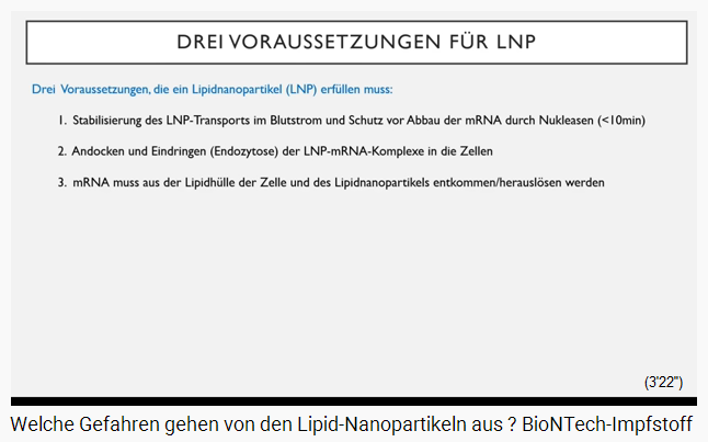
SLIDE 2: The properties that lipid nanoparticles (LNP) must have
(orig. German: FOLIE 2: Die Eigenschaften, die Lipidnanopartikel (LNP) haben müssen)Title of the text panel: Three requirements for LNP:
Three requirements that a lipid nanoparticle (LNP) must meet:
1. Stabilization of LNP transport in the bloodstream and protection against degradation of the mRNA by nucleases (<10min)
2. Docking and penetration (endocytosis) of the LNP-mRNA complexes into the cells
3. mRNA must escape / be dissolved out of the lipid envelope of the cell and of the lipid nanoparticle (3'12 '')
So: The lipid nanoparticles that are manufactured must meet the [following] requirements (3'18 ") so that the technology can work (3'20").
Point 1: Lipids that are used must ensure the transport of nanoparticles in the bloodstream (3'28 '') and also for the protection of the degradation of the mRNA, this was mentioned at the beginning already (3'35 '').
Point 2: These lipid nanoparticles must of course also be able to dock on the respective body cells (3'43 ''), and also be absorbed by them (3'46 ''), so this penetration is called endridosis [?] - technical term (3'50 ''), so that the mRNA gets into the cell at all (3'53 '').
[Point 3]. And as a third point, the mRNA has to get out of this lipid envelope, but not only from the lipid envelope of the cell, but also from the lipid envelope of the nanoparticle (4'5 '').
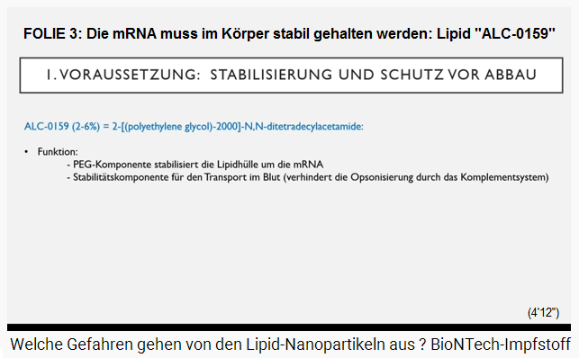
SLIDE 3: The mRNA must be kept stable in the body: Lipid "ALC-0159"
(original German: FOLIE 3: Die mRNA muss im Körper stabil gehalten werden: Lipid "ALC-0159")
Title: 1. REQUIREMENT: STABILIZATION AND PROTECTION AGAINST DEGRADATION
ALC-0159 (2-6%) = 2 - [(polyethylene glycol) -2000] -N.N-ditetradecylacetamide:
-- Function:
-- the PEG component stabilizes the lipid envelope around the mRNA
-- Stability component for transport in the blood (prevents opsonization by the complement system) (4'6 '')
The lipid: ALC-0159 = 2 - [(polyethylene glycol) -2000] -N, N-ditetradecylacetamide [PEG-2000]
Let us first come to the prerequisite, namely the stabilization and protection against degradation (4'12 ''), lipid with the name ALC-0159 is doing this (4'17 '') a 6% rate is in it, and that is the typical - [now I need a] pen - that's the typical PEG (polyethylene glycol) (4'27 ''). But there is more to it here, it is not pure PEG (4'31 ''), but it has another, another molecule to it (4'34 ''), it is a - let's call it a "linker" (NN-ditetradecylacetamide), that is important later when explaining, yes, how the whole process works (4'43 '').
So this lipid is there on the one hand to stabilize the lipid envelope (4'50 ''), and secondly for the stability of the transport in the blood (4'57 ''), because this PEG is like that protective layer around this lipid nanoparticle (5'6 ''), so that the complement system - that is a part of the innate immune system (5'11 '') - consists of many different proteins (5'14 '') , does not recognize this lipid nanoparticle (5'18 ''), because if it were to recognize it, then it would lie around it and mark it, so to speak, for macrophages [for the white blood cells] that are in a good mood (5'27 ''). Macrophages [white blood cells] I already explained to you in the first video, are always the vacuum cleaners of the blood (5'32 ''), they recognize foreign bodies and pick them up and break them down (5'36 ''). And this process is called optionization [?] (5'39 ''). And for blocking this, there is PEG, so these particles in the blood are in principle made a bit inconspicuous (5'49 '') so that they are not degraded down immediately (5'51 '').
The second requirement is the fusion and penetration of the lipid nanoparticle with the mRNA into the cell (6'2 '').
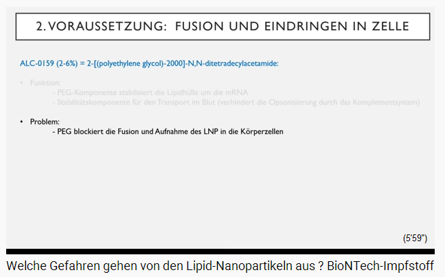
SLIDE 4a. Precondition: fusion and penetration into cell:
[Lipid]: ALC-0159 (2-6%) = 2 - [(polyethylene glycol) -2000] -N.N-ditetradecylacetamide [this is: PEG-2000]:
Problem:
-- PEG blocks the fusion and uptake of the LNP in the body cell
Um, yes: The problem when you have PEG - so without PEG the whole system doesn't work because the particle is broken down immediately (6'13 '') - it's unstable, but when the PEG is inside, it blocks it at the same time also the docking and the fusion of the nanoparticle in the body cell pure (6'22 '').
The problem can be solved by including cholesterol in this nanoparticle (6'31 '').
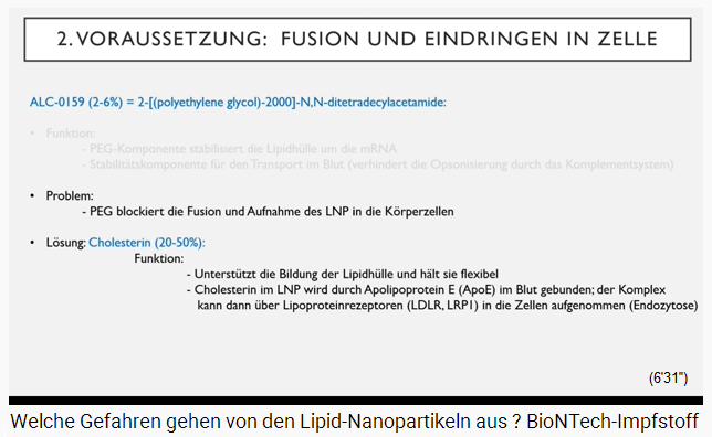
SLIDE 4b. Cholesterol attached at the mRNA vesicle
Precondition: fusion and penetration into cell:
[Lipid]: ALC-0159 (2-6%) = 2 - [(polyethylene glycol) -2000] -N.N-ditetradecylacetamide [PEG-2000]
Problem:
-- PEG blocks the fusion and uptake of the LNP in the body cell
Solution: cholesterol (20-50%):
Function:
-- Supports the formation of the lipid envelope and keeps it flexible
-- Cholesterol in the LNP is bound by apolipoprotein E (ApoE) in the blood: the complex can then be absorbed into the cells via lipoprotein receptors (LDLR, LRPI) (endocytosis) (6'26 '')
Cholesterol is about 20 to 50% in the BionTech vaccine [Pfizer vaccine] (6'38 "), and the function of cholesterol is primarily: It supports the formation of the lipid envelope and keeps it flexible (6'45 '') that the lipids can swim freely a bit (6'48 ''), and the second function is: The cholesterol is bound by apolipoprotein in the blood (6'56 ''), so apolipoprotein E is a molecule that floats in the blood (7'0 '') and finds cholesterol (7'3 ''), namely the cholesterol on the lipid envelope of this particle (7'7 ''). And when it finds that, it binds this cholesterol (7'13 ''), and this complex via apolipoprotein E is recognized by the cells (7'19 ''), and these cells have lipoprotein receptors (7'23 ''), there are a number of receptors in nature (7'26 ''). The best known are the LDL receptor (7'29 '') and the LMP1 (7'31 ''). And these receptors bind apolipoprotein E, which in turn is bound to cholesterol (7'39 ''), which in turn carries the lipid nanoparticle (7'41 ''). And when this whole complex binds with each other, then this complex is actively incorporated by the cell, and the whole thing is then inside the cell at once (7'54 '').
Now we come to the third requirement (7'59 ''): Now the mRNA still has to be freed from the shell (8'2 ''). I've listed a few things here (8'5 ''):
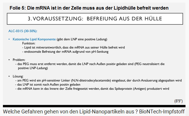
SLIDE 5: The mRNA is in the cell and must now be freed from the lipid envelope
(original German: FOLIE 5: Die mRNA ist in der Zelle drin und muss nun aus der Lipidhülle befreit werden)
Precondition: release from the shellSo primarily the lipid ALC-0315 is responsible for it (8'16 ''). A rate of 30 to 50% is available in the BionTech [Pfizer poison injection] in a fabric lipid shell (8'20 ''). That is a cationic area (8'23 ''). What does cationic mean? These are all molecules that migrate to the cathode (8'27 ''), so when you were paying attention in your chemistry lessons: The cathode is the pole that is negative (8'34 ''), i.e. molecules that are positively charged, are migrating to the cathode (8'39 ''). That means: The cationic lipid is positively charged (8'41 ''). It gives, so to speak, a positive, electrical charge to the whole lipid nanoparticle (8'49 ''). So, this lipid is partly responsible for the fact that the mRNA is freed from a shell (8'53 ''), and this finally happens via a pH decrease that is proceeded in the cell (8'59 ''). This is all complicated, therefore I am going on straight to the next slide (9'2 ''), where I'll draw it again, because then it's much easier to understand it (9'6 '').
[Lipid]: ALC-0315 (30-50%):
-- Cationic lipid component (gives the lipid nanoparticle LNP a positive charge)
Function:
-- the lipid is partly responsible for the fact that the mRNA is released from its shell
-- endosomal liberation of the mRNA by lowering the pH
Problem:
-- the polyethylene glycol (PEG) must first be removed so that the lipid nanoparticles LNP are positively charged to the outside (PEG neutralizes the positive LNP charge)
Solution:
-- A pH-sensitive linker (N.N-ditetradecylacetamide) is integrated into the PEG, which is split off by acidification
-- the lipid nanoparticle (LNP) is thus positively charged to the outside - the mRNA can be released into the interior of the cell so that the spike protein (antigen) is produced
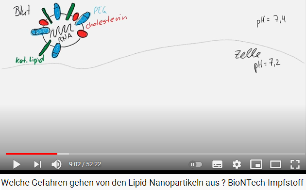
SLIDE 6a (drawing): Drawing of the spike protein in a body cell, pH values 7.2 and 7.4
(original German: FOLIE 6a (Zeichnung): Zeichnung des Spike-Proteins bei einer Körperzelle, pH-Werte 7,2 und 7,4)
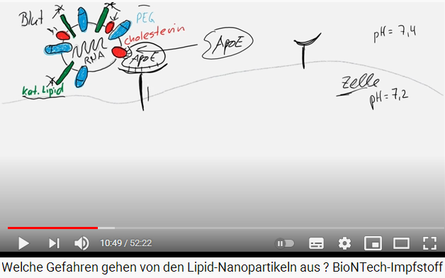
Let's take a pen again (9'11 ''): So, at the top left you can see the lipid nanoparticles (9'14 ''), in the middle the mRNA (9'17 ''), I've already marked you in color the various components that are in there (9'19 ''), on the one hand the cationic lipid in green (9'26 ''), then we have the polyethylene glycol (PEG) [in light blue] (9'30 ''). Well, the cationic lipid, I have to say again, has a positive charge on the outside (9'37 ''), and the PEG, which stabilizes the whole complex (9'41 ''). The problem with PEG, however, is that it inhibits this positive charge, which means that ultimately this lipid nanoparticle is neutral again (9'53 ''). It is important to say now, because later this cationic lipid has a function (9'58 ''). At the moment this nanoparticle still has a neutral charge (10'3 ''). So, and then there is the cholesterol, which I chose here in red (10'6 ''). So, and APOE molecules are now swimming in the blood (10'12''). And THESE find the cholesterol (10'15 ''), and they bind to the cholesterol (10'17 ''). Here an interaction is taking place (10'21 ''). It also takes place here, or here [wherever cholesterol surrounds the mRNA]. (10'25 ''). Several APOE molecules can naturally bind the lipid (10'29 ''). And on the cell surface here - that's the cell (10'32 '') - below and above is the blood (10'35 '') - on the cell surface there are receptors (10'37 '') - I had called, these are the apolipoprotein receptors (10'43 '). And they recognize the APOE (10'47 ''). And they also bind here (10'49 '').
SLIDE 6b (drawing): drawing of the spike protein with reactions with APOE molecule and apolipo protein etc.
(original German: FOLIE 6b (Zeichnung): Zeichnung des Spike-Proteins mit Reaktionen mit APOE-Molekül und Apolipo-Protein etc.)
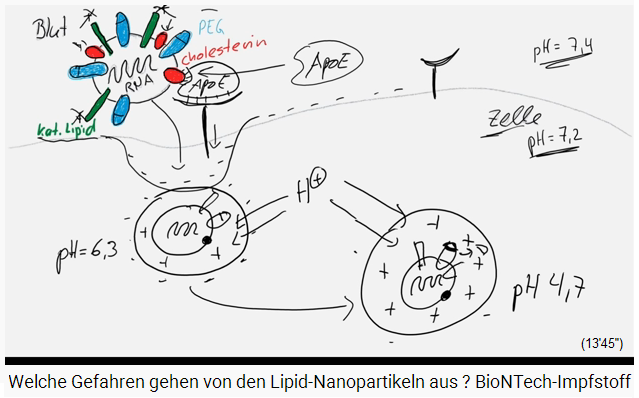
And now all this emits a signal in the cell that the whole thing is now being incorporated (10'55 ''). And then we basically have the mRNA with this lipid shell [with all these "spikes"] (11'9 "), and since there is such an indentation in the cell membrane (11'13"), we have the envelope of the cell around it (11'17 ''), this is another lipid envelope (11'18 ''). So, and this cell membrane has a negative charge, because the lipids - that becomes quite chemical now, but that is important to understand the side effects afterwards (11'31 ''), because these lipids have a head that is negatively charged (11'36 "), that means: Here we have everywhere negative charges [she is drawing points around the rims ] (11'41"), and what happens now is a normal, a normal, natural process (11 '47' '), whatever happens in a cell, if the cell absorbs something (11'49' '), for example a hormone or insulin for example (11'53' '), then it happens that the content which was incorporated from the outside world has to be released somehow (12'1 ''). There are molecules for that - I just call them molecules now (12'6 ''), they manage to pump protons from the outside into a cell, but from the cytodol [?] of the cell, into this vesicle (12 ' 21 ''). Therefore now, proton [?] is accumulating in the vesicle and that means the pH value goes down (12'30 ''). So, here outside we still have a pH value of 7.2, in the blood around 7.4, and here in the vesicle at the beginning we already have a pH value of 6.3 (12'43 '').
SLIDE 6c (drawing): The spike protein is in the cell with a pH of 6.3
(original German: FOLIE 6c (Zeichnung): Das Spike-Protein ist in der Zelle mit einem pH-Wert von 6,3)
And that takes a while, more and more protons come in (12'49 ''), and then there is - then it goes so far down to a pH value of about 4.7 (13'0 ''), I think that's the maximum (13'3 ''). So more and more protons are entering (13'6 ''), so, draw it, that's important (13'17 ''), the negative charge that goes away here (13'19 ''), it's only weak yet (13'21 ''), we have a lot of positive [charge] in here (13'23 ''), and this negative pH value, not negative, so this small [low] acidic pH value causes that this linker is split off from this PEG lipid here above [drawing: one end of a cholesterol [oval + red] is marked and provided with an arrow pointing outwards] (13'40 ''). That means there is still something left (13'45 '').
SLIDE 6d (drawing): The vesicle has pH 4.7 - is positively charged and the cap of the cholesterol is gone
(original German: FOLIE 6d (Zeichnung): Das Vesikel hat pH-Wert 4,7 - ist positiv aufgeladen und die Kappe des Cholesterins ist weg)
Slide 6d (drawing): The vesicle has pH 4.7, is positively charged and the cholesterol cap was split off [6d]
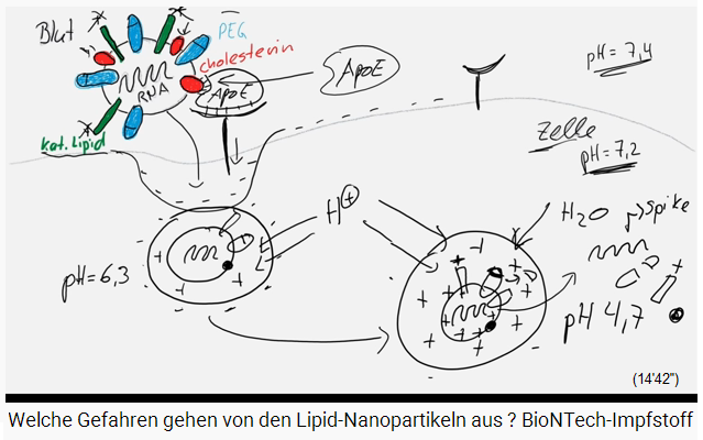
And when it [the "linker"] is split off, it no longer inhibits [blocks] the charge from the cationic lipid (13'51 ''). That means: The charge of the lipid nanoparticle is also positive (13'58 ''). And if everything is positive now - that is of course much more positive than here in the cell (14'6 ''), then of course water flows in (H2O) (14'9 ''), without end (14'11 ' '). And like a balloon with too much air in it, this vesicle bursts (14'18 ''), and when it bursts, of course the mRNA is released, it can then produce the spike protein (14'27 '' ), but also the other components, here the linker, the polyethylene glycol (PEG), the cationic peptide (14'36 ''), cholesterol, everything is released (14'38 ''). That is now all in the room of the cell (14'42 ''),
SLIDE 6e (drawing): The vesicle with the influx of water bursts and the mRNA and lipids are free in the cell
(original German: FOLIE 6e (Zeichnung): Das Vesikel mit Wasserzustrom platzt und die mRNA und die Lipide sind frei in der Zelle)
Slide 6e (drawing): The vesicle bursts with the influx of water and the mRNA and lipids are free in the cell [6e]
What is very important for later: This here, the cationic lipid, really provokes difficulties causing massive cell toxicity (14'53 ''), so, it is toxic for the cells (14'54 ''). First and foremost, of course, the mRNA, not cholesterol, the PEG itself is not harmful either (15'1 ''), but the cationic lipid (15'4 ''). So you have to remember two things: The cationic lipid is very harmful (15'8 ''), and the whole thing is absorbed by APOE and APOE receptors, which are located on the cell surface (15'15 '').
SLIDE 6f (drawing): The burst vesicle in the cell: Toxic substances are marked in a circle: the cationic lipid and the mRNA
(original German: FOLIE 6f (Zeichnung): Das geplatzte Vesikel in der Zelle: Giftige Substanzen sind eingekreist markiert: Das kationische Lipid und die mRNA
Where do the lipid nanoparticles actually go? (15'23 '')
Slide 6f (drawing): The burst vesicle in the cell: Toxic substances are marked in a circle: The cationic lipid and the mRNA [6f]
SLIDE 7a + b: Where do the lipid nanoparticles go?
(original German: FOLIE 7a+b: Wo gehen die Lipidnanopartikel hin? )
Tests on mice and rats: Detection of lipid nanoparticles (LNP) within 1 hour after the injection: 1. At the location of the puncture site 2. In the blood 3. Transport via the blood to other organs: - bone marrow - liver - lungs - spleen - kidney .Where do the lipid nanoparticles really go? (15'23 '') - I mean, we get the injection in the upper arm, in the muscle (15'28 ''), everyone would think: Yes, well, then it's there on site (15'32 '') . But there are experiments on mice and rats, so these experiments have been going on for over 10 years, I have to say (15'38 ''), and then one could see clearly, and this came also out clearly in many publications, in all I read (15'45 '') that the lipid nanoparticles are not only at the site of the puncture site (15'50 ''), but they are also found relatively quickly in the blood (15'54 ''), and with the blood they can of course be transported to all organs (15'59 ''). And you can find them especially - well, it's definitely published - [now let's look at the list]: in the bone marrow (16'6 ''), in the liver, in the lungs, in the spleen, in the kidney (16 ' 10 ''), especially liver lungs spleen were affected (16'12 ''), the duration of the detection was now 10 days (16'14 ''), depending on how fast the metabolism is (16'17''), mice and rats have faster metabolism and turnover, for us humans it will certainly take a little longer, I think (16'23 ''), but it depends on how much is injected (16'25 ''). In addition, there is no data for it yet (16'28 ''). I have now inserted a few pictures for you, what it looks like (16'36 ").
Duration of proof: 10 days. Tests on humans (preclinical): lipid nanoparticles (LNP) were found in all tissues: especially in spleen and liver.
BioNTech / Pfizer Public Assessment Report (Study R-20-0072 and PF-07302048):
Experiments on rats with lipid nanoparticles LNP (same composition as in the vaccine) + luciferase mRNA:
After 2 weeks and after only one injection, they could still detect the luciferase activity in the blood and in the liver, as well as at the injection site (other organs are not mentioned). After the intramuscular injection, the luciferase activity was already detected 6 hours later in the tissue at the puncture site and in the liver (=> simultaneous production of luciferase) (15'21 '').
The photo above with the liver: on the photo above there is a part of the liver, this is a microscopic photo (16'40"), in the green [area] is the cell membrane painted on by the different cells (16'42 ''), what you can see here are the individual liver cells (16'46 ''), so here one, here one, here one, these are the individual liver cells (16'52 ''). and the pink (violet), that is the dye that has been packaged in the lipid particles (17'0 ''), the pink are basically the places where the nanoparticles have come to (17'5 ''), and this can be seen very well: the complete tissue of the liver is full of these lipid nanoparticles (17'13'').
The right photo with the spleen: Now see the photo with the spleen: here is the spleen, and on the rim one can see - well, this is this zone here [she marks a red half moon], this is the Red Pulp of the spleen, this is called like this (17'25''), this is the zone which is especially supplied with blood (17'28''), there are the macrophages [white blood cells], these are the vacuum cleaners of the blood (17'31") waiting there, the blood is passing there and they [the white blood cells] are looking for and taking out all the foreign bodies (17'37 '').
Slide 7b: Where do the lipid nanoparticles go? The lipid nanoparticles (colored in purple) mainly affect liver and spleen (with markings) [7b]
This is a kind of purification of the blood, one can say (17'41 ''), but with spleen it's absolutely sure (17'44''), well, here in the middle, this is the white substance of the spleen (17'47''), this has another function than the red substance (17'51''). But also here: the red substance is massively attacked by the lipid nanoparticles (17'56'').
The photo with the complete rat: Then we have here in the middle an X-ray photo (18'2''), the lipid nanoparticles have been marked with an offset dye on the top (18'6''), the bottom has been radioactively marked (18'9 ' '), and you can see very well: the darker the structure, the more the tissue is infected (18'16' ') with lipid nanoparticles (18'18' '), and you can see very clearly here that the spleen and the liver is affected here (18'21 ''). The lungs are affected, the kidneys (18'25 ''), but the method is not particularly sensitive, that's why you don't see it so well, you actually have to cut open to see it better (18'32 '') that the lipid nanoparticles do NOT linger at the puncture site, but spread very quickly over the entire tissue (18'41 ''). So, then there are experiments on humans, the preclinical (18'44 ''), where it was also found that these lipid nanoparticles were found in all tissues (18'49 ''), but especially in the spleen and in the liver (18'52 '').
So, then there is the Public Assessment Report from BionTech / Pfizer (18'58'') the study 20 72 (R-20-0072) and the strain PF-07302048 (19'3 ''), they proceeded a trial with rats (19'5''), and they were injecting nanoparticles, the composition of these lipids was the same as with the vaccine they are using with us (19'13''), but in the middle was not the mRNA2-proteine, but there was an mRNA luciferase (19'19''), this is a [not understandable] (19'22''). And after 2 weeks - just after ONE injection - one could prove activity of the luciferase in the blood an in the liver yet (19'28''), with animals as also at the injection site (19'32''), and other organs were not at all mentioned, were - perhaps they were not at all investigated (19'37''). Uhm, after the intermuscular injection - that's basically the same method that you use with us, injecting it into the muscle (19'45 '') - this luciferase activity was found after 6 hours after the injection already in the tissue of the injection zone (19'53''), abut also in the liver it could be found 6 hours after the injection already (19'59''), this means: the distribution of the nanoparticles is very fast (20'3''). Well, being injected into the muscle, so this will be pretty fast in the liver (20'10''). And the production of the spike protein will be proceeded practically at the same time in all organs (20'16'').
SLIDE 8: Rats injected 3 times in 2 weeks with gene "vaccination" from Pfizer - and the immune response
(original German: FOLIE 8: Ratten 3mal in 2 Wochen mit Gen-"Impfung" von Pfizer gespritzt - und die Immunantwort)
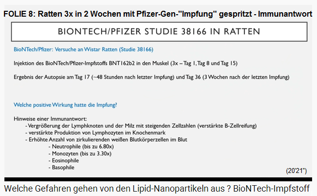
Now Biontech / Pfizer has done another study, the study 38166 in rats, Wistar rats (20'24 ''), and they have not taken the luciferase, but they really took the vaccine which is also used with us, the real one (20'31''), and this vaccine was injected the rats into their muscle (20'36''), [all in all] three times , at day number 1, number 8 and number 15 (20'40''). And then 2 days after the last injection the took one part of the rats proceeding an autopsy, and 3 weeks after the last injection the other part of the rats was killed for performing an autopsy (20'51'').
So what positive effects did the vaccination have?
Study Biontech / Pfizer Study No. 38166 with rats:
BioNTech / Pfizer: Tests on Wistar rats (Study 38166)
Injection of the BionTech vaccine BNT162b2 into the muscle (3x day 1, day 8 and day 15)
Result of the autopsy on day 17 (until 48 hours after the last vaccination) and day 36 (3 weeks after the last vaccination)
-- What positive effects did the vaccination have?
-- Indications of an immune response:
-- Enlargement of the lymph nodes and the spleen with increasing cell numbers (increased B-cell maturation)
-- increased production of lymphocytes in the bone marrow
-- Increased number of circulating white blood cells in the blood
-- Neutrophils (up to 6.80x)
-- monocytes (up to 3.30x)
-- eosinophils
-- Basophils (20'22 '')The immune response: Increased production of lymphocytes in the bone marrow - migration to the spleen - maturation and enlarged spleen + lymph nodes
SLIDE 8: Rats get 3 injections within 2 weeks with the genetic Pfizer "vaccine" - - and the immune response [8]
In THIS study they analyzed a little DEEPER (20'57 ''). So there is - they say, there is already evidence of an immune response (21'2 ''), there is an "increased production of lymphocytes in the bone marrow" (21'10 ''), lymphocytes reproduce themselves - dividing themselves and reproducing themselves and they are created in the marrow (21'16''), and then they migrate from the marrow (21'20'') migrating to the spleen where they are maturing principally (21'24''). Also there they remain and when they are needed they mature to be B cells for example (21'30''). Well, one can see more production of lymphocytes, these are the immune cells of the marrow (21'35''), and at the same time, more of these cells are maturing in the spleen (21'39''), because the spleen, well, they were counting the cell days [?], but this spleen is enlarged then (21'45''), as are also the lymph nodes enlarged (21'47'').
Immune response: More white blood cells in the blood
Then they also found an increased number of circulating white blood cells in the blood (21'52 ''), which is evidence of an immune response (21'57 '').
Now we come to the side effects that were: So, they wrote (22'3 ''):
-- The rats had an increased body temperature (22'5 '')
-- they also had a body weight loss (22'8
And body weight loss always means, well, this is the case with rodents (22'13''), that they loose their weight fast when they suffer a stress situation (22'16''). This does not only mean psychical stress, but really also physical stress in the body (22'19''), thus when their body is not working well, then they are loosing weight very fast (22'24''), and this is with the rodents, this is - this is really a factor where one can say: the mouse or the rat is not doing well (22'34'').
The inflammatory reaction at the puncture site: instead of muscle cells, there is only incrustation (connective tissue) - the muscle's function is limited
So now, there is a local inflammatory reaction at the puncture site (22'39''), and this inflammation is infection the whole zone around it (22'44''). Skin becomes red and swollen (22'47''). This is principally the same what was told in the Clinic study 1, what we can see when there is an injection in the muscle - well - when there is an injection given there, the reaction was always a swollen zone and tissue [?] and a red skin, this can be seen with the rats, too (23'3''). But what cannot be seen with us is, but this COULD BE SEEN with the rats because they were killed with an autopsy then (23'12''): tissue was taken out and was analyzed then (23'15''), and it was shown that they had a hardened tissue, [special word] incrustation (23'20''). Incrustation means: salts are incorporated in the necrotic [dead] tissue (23'24''). Well, this necrotic tissue is finally tissue which is dying (23'27''). This means: With an injection at a certain point where it was put in (23'33'') the tissue has died or is dying yet depending on the time of procedure (23'39''). Another indication of it is the incorporation of fibrosis, this is a reproduction of connective tissue in a pathological increase (23'47''), normally this is called fibrosis (23'50'').
Ultimately, that means: Collagen fibers are stored (23'53 ''), the tissue that has died up here, what has died here, has to be replaced somehow. This is then replaced by connective tissue (23'59 ''). This connective tissue consists of cells that are different from the cells that were there before (24'3 ''). So: Before it was muscle cells, that is not there now, because now there are these fibril-like cells (24'10 ''), and of course they have a completely different function: This restricts the function of the actual organ, namely the muscle, so , the function is restricted (24'19 ''). So, well, they said that they also saw the death of the muscle fiber cells on a massive scale (24'27 '').
THIS is principally just what I feared of, and what I was stating in my first video (24'33''), that is that these lipid nanoparticles are simply toxic (24'36''). That means: If I throw these lipid nanoparticles on my cell culture in the laboratory and I have put too much of them (24'46 ''), they are dead the next day. My cells [of the cell culture] then no longer exist (24 ' 50 ''). And exactly the same thing apparently happened here with the rats (24'52 ''). The muscle fibers died (24'57 ''), it didn't look good - that came to the necrotic tissue and instead of it a connective tissue came that we don't want at all (25'3 '') . And they also say here that this phenotype became particularly severe and was seen more often (25'13 '') because after the second or third vaccination, so, the more often you inject the poison into the rats, the worse it was (25'22 ''). Then they wrote yet that the increase in proteins in the blood was detectable (25'27 ''), for example the first one here: Fibrinogen up to 2.49 times as much [as normal] (25'34 ''), fibrinogen is an indication of an inflammation of the blood vessels (25'39 ''). It has the task of blood clotting (25'42 '').
The muscle cells: 13 different cell types
Yes, now you also have to know: The muscle consists not only of muscle cells (25'48''), well, muscle fibers have two different types, but a new study shows that there exist about 13 different cell types in the muscle (25'55''). Of course, there are stem cells which are developing forming muscle again 826'0''), there are different immune cells which have to maintain the muscle healthy (26'4''), there are endothelial cells which are principally creating blood vessels (26'10''), there are pericytes which are around T cells (26'13''), which are let's say one of the factors responsible for the contraction of the blood vessels (26'16''), there are many different cell types [in the muscle] (26'20'')
The lipid nanoparticles destroy cell walls - blood vessel ruptures occur - bleedings occur - and the fibrinogen wants to clot the flowing blood
And the lipid nanoparticles, which are toxic of course (26'28 ''). Some cells are sensitive, others less sensitive (26'32''), but obviously there exist little blood vessel ruptures here (26'37''), this means that the lipid nanoparticles also have invaded the NOT cells and destroyed them (26'43''), otherwise there would not exist slight bleeding there (26'46''), and [otherwise] there would not be fibrinogen synthesized which has got the task to clot the flowing blood at the same spot again (26'53''). Otherwise we would die by bleeding when it becomes too bulky (26'56'').
Other indicators of inflammation: alpha-2 macroglobulin and AGP
So then we have the second protein, alpha-2 macroglobulin (27'2 "), which is 71 times increased (27'4"), which is also part of the immune response, which is caused by inflammation [not understandable] (27'9 ''). The third is AGP, which has been increased up to 39 times (27'15 ''), and this is also produced when there is increased tissue damage in the body (27'21 ''), which is the case with inflammation and also with infections (27'24 '').
The tissue is destroyed after just 3 weeks - the culprit is lipid nanoparticles
So, all these schemes that we see here, that is absolutely something that, in my opinion, comes about through these lipid-nanoparticle complexes, and not because of any immune response (27'39 '') that may begin after 3 or 4 weeks in the used rats (27'42 '') because: The spike protein - well, the first rats were analyzed after 3 weeks already (27'50 ''), there aren't any real antibodies yet in large amounts (27'53 ''). And these are all - this phenotype, these are all indications that the lipid nanoparticles, i.e. the lipids themselves, massively destroy the tissue (28'6 '').
I show you a picture now of where exactly you can find the lipid nanoparticles in the liver (28'17 '').
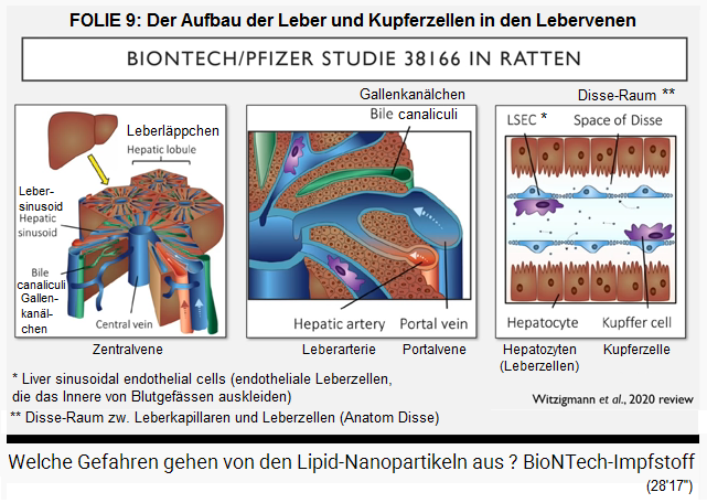
SLIDE 9: The structure of the liver - the copper cell in the hepatic vein
(original German: FOLIE 9: Der Aufbau der Leber - die Kupferzelle in der Lebervene)
Up here, just a moment please, this is the liver which was cut in a cross-section (28'27''), and then one can see the liver modules, these are no cells, but complete modules (28'32''). Well, blue are the veins (28'34''), red are the arteries (28'37''), and the brown mass are the liver cells (28'39''). And zooming in one can find this vein, the portal vein, and by this vein the lipid nanoparticles are coming into the liver (28'55''), and now showing the next zoom these are liver cells (28'58''). The brown cells are the liver cells (29'1''). And zooming again one can see the scheme: lipid nanoparticles are coming in (29'11''), they are meeting rose purple cupper cells (29'16''), they are marked in purple dye, these are the hoovers in the liver, the white blood cells marcophages (29'22''), there is blood and there are copper cells in the liver (29'25''). And nano particles were found here massively in the cupper cells (29'32''), they are trying to shift away the garbage, to remove it from the liver (29'38'') because the lipid nanoparticles are a foreigner for the liver (29'41''). Lipid nanoparticles are present here also because, this was found out, massively all over here in the liver cells (hepatocytes) (29'53''). This means, see again the photo of before (29'55''), with the pinky dye (29'58''), these lipid nanoparticles were invading the liver here over all (30'3'').
The damaged liver cells: vacuolation (the liver becomes holey like a Swiss Emmental cheese)
So, and I told you, in the muscle, what devastating destruction these lipid nanoparticles do (30'13 ''). So of course the same thing happens in the liver (30'17 ''). The liver is damaged (30'19 ''). And I took a picture of what it could look like down here (30'23 '').
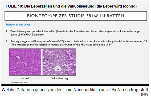
SLIDE 10: The liver cells and the vacuolation (the liver becomes holey like a Swiss Emmental cheese)
(original German: FOLIE 10: Die Leberzellen und die Vakuolisierung (die Leber wird löchrig wie ein Emmentaler Käse)
Slide 10: Damage to the Liver:
1. Vacuolization of portal liver cells (evidence of the death of liver cells due to liver injuries caused by lipid nanoparticles LNP / mRNA complexes)
2. Increase in gamma-glutamyltransferase (GGT) - various causes: Liver disease due to drugs or poison "The vacuolation may be related to hepatic distribution of the PEGylated lipid in the LNP" (30'5 '').
The photo here on the left side, this is a cross-section of the liver tissue in a normal, healthy state (30'33''), one can see here overall the singular cells (30'37''), here inside these are two blood vessels (30'40''), and the pink dye is finally glycogen, glycogen was colored, this is the sugar, it is stored in form of glycogen in the liver as an energy deposit (30'53''). And here on the right photo one can see a vacuolation (30'56''). This vacuolation here above - they told in the BionTech study with rats - they said:
"Vacuolization takes place in the portal liver cells" (31'8 ''), and vacuolization always forms - that is the proof that the liver cells die, namely: They die due to liver injuries (31'17 ' '). And they could only have got this injury through these lipid nanoparticles (31'21 '').
(original German:See the photo again, these here are vacuoles [liver cells] (31'28''). There are simply holes where were liver cells before, they have gone now (31'32''), dreadful, the complete tissue [of the liver] is holey [like a Swiss Emmental cheese] (31'35''). Here is a blood vessel which is no longer correct (31'37''), then here is pinky color which is less strong [is less intensive] because glycogen cannot be stored as much as before when the liver cells loose their function 831'46''). Yes, and this yet, just by the way, this is also the kind of liver of an alcoholic or of a person with extreme overweight (31'58''). Yes, this is also called dystiatosis (32'2'').
"Es findet in den portalen Leberzellen eine Vakuolisierung statt" (31'8''), und eine Vakuolisierung bildet immer - das ist der Beweis dafür, dass die Leberzellen absterben, und zwar: Sie sterben ab aufgrund von Leberverletzungen (31'17''). Und diese Verletzung können sie nur durch diese Lipidnanopartikel gekriegt haben (31'21'').
We have seen that there is an increase in GDT (32'10 ''), this is an enzyme in the liver (32'13 ''), many people know this value if you do a normal blood analysis at the doctor's, because the various liver values are also tested (32'22 ''), and then also the GDT (32'24 ''). If it is too high, then of course that is not good, there are various reasons for it (32'30 ''):
-- One cause of this is a liver disease caused by a medicament or a toxic poison (32'35 '').
And even BionTech writes - we go down to this sentence - that is a quote from their publication (32'45 '') that they suspect that vacuolization takes place in these liver cells due to the PEG lipid in their lipid nanoparticles (32'55 ' ').
So they know very well that their stuff damages the liver (33'2 ''). Why isn't it mentioned in the media? Incredible! (33'6 '')
Personally, I think it's not the PEG, the PEG alone is not toxic to humans (33'12 ''). I think it's the cationic lipid (33'14 ''). Later I will explain why this is so (33'17 '').
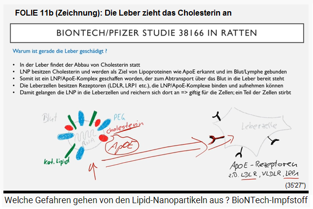
Cholesterol is degraded down in the liver - that's why there is a liver damage
So why occurs this damage just in the liver? (33'23'') Here is the lipid, a nanoparticle is just a composition of different lipid components (33'34''), and I told you about cholesterol that it binds APOE in the blood (33'42''). And this APOE in the blood basically - well, the liver has got a function, has got many functions, but one of the principle functions of the liver is the degradation of cholesterol (33'54''). well, degradation of cholesterol is proceeded in the liver (33'57''). And this APOE binds cholesterol and is only doing the transportation of the cholesterol to the liver by swimming in the blood (34'9'').
is the liver damaged? (33'23 '') Here is this lipid, nanoparticles are a compilation of the various lipid components (33'34 ''), and I told you about cholesterol that binds this APOE in the blood (33'42 '' ). And this APOE in the blood has basically - so the liver has one, has many functions, but has one main function, for example it is also the breakdown of cholesterol (33'54 ''). Well, the breakdown of cholesterol takes place in the liver (33'57 ''). And this APOE binds cholesterol and has nothing more to do than to bring the cholesterol that it has bound back to the liver via the blood (34'9 '').
SLIDE 11a: Graphic of the spike protein with cholesterol, and a liver cell with receptors for the cholesterol
(original German: FOLIE 11a: Grafik des Spike-Proteins mit Cholesterin, und eine Leberzelle mit Rezeptoren für das Cholesterin)
SLIDE 11a: Graphic of the spike protein with cholesterol, and a liver cell with receptors for cholesterol [11a]
Well, this is what it knows: Binding something, depends on what - the whole thing is a little bit more complicated, but now this is a rough presentation (34'16'') - when the APOE is binding the cholesterol in a complex, then it will migrate back to the liver [where is is degraded] being sucked by the APOE receptors (34'28'')
which I mentioned at the beginning, even if it also has the muscle cell, almost actually, I don't know of any cell that doesn't have that (34'35 '') - there are so many different APOE receptors (34'37 '' ), every cell will have something (34'39 ''). But the liver in particular has LDL receptors and P1, and by this it is incorporated into the liver cell (34'48''). Well, now the liver cells are sucking all the cholesterol concentrating it in the liver cells because this APOE - this lipid nanoparticle is bound to the cholesterol, and it comes in, too, with full intention (35'3''). With other tissues it is not such an intention (35'6'').
So, the lipid nanoparticle is passing a cell, and when it's passing a receptor by the way it is sucked and integrated (35'15''). But this is really a transport, this is a normal mechanism (35'19''), how cholesterol is eliminated from the blood, how the lipid metabolism is working in humans (35'24''), well, and therefore the liver is especially affected by the damages [cell damages by lipids], this is caused by the vaccination (35'32'').
So there this lipid nanoparticle simply runs past a cell, and if it happens to have such a receptor, then it is absorbed (35'15 ''). But that is really a transport here, that is a completely normal mechanism (35'19 ''), how cholesterol is removed from the blood, how the lipid metabolism works in humans (35'24 ''), yes, and therefore the liver will be particularly badly affected by the damage [cell damage by lipids] caused by this vaccination (35'32 '').
After Pfizer gene vaccination: Lymphocyte levels are low for 1 week - lymphopenia - people who have been vaccinated should be quarantined for 1 week
Well, then I also explained or showed in my first video that in the Clinic study 1 there can be a massive decrease in lymphocytes in the test subjects (35'47 ''), already after ONE day after the first vaccination ( 35'49 ''). This decrease in lymphocytes was normalized again after one week (35'54 ''),
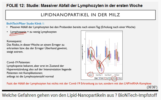
SLIDE 12: Massive decrease in lymphocytes in the first week
(orig. German: FOLIE 12: Studie: Massiver Abfall der Lymphozyten in der ersten Woche)
Slide: LIPID NANOPARTICLE IN THE SPLEEN
BionTech / Pfizer Study Clinic 1:
-- Lymphocytes are massively decreasing in the test subjects after just one day (recovery after one week)
-- Lymphopenia = too few lymphocytes
Consequence:
The risk of getting an infection during this week resp. that a pathogen will will gain the upper hand increases extremely.
Covid-19 patients: lymphopenia is known, but only in a state of hyperinflammation; that is, patients in the intensive care unit with complications; initially the number of lymphocytes is normal.
Conclusion: The decrease in lymphocytes has nothing to do with Covid 19 disease, but with the LNP / mRNA complexes (35'36 '')
So there was some kind of recovery (35'56 ''). You can see that here [on the graphic]: Well here, this is the base line [first group of values], these are the vaccination samples [second group of values] (36'2 ''), and depending on the concentration and with us again 30 micrograms, so we would have to look at the brown [look at the brown line] (36'10 ''), from here to here there is a decrease [falling level] of the lymphocytes (36'13 ''), and after one week it's up again (36'15 '').
So, when a person has got too few lymphocytes in the blood, this is defined as a lymphopenia, this I noted above (36'25''), here of course this is not the case, so there is no illness basically (36'30''), but I only wanted to teach you this special term because many persons have such a problem (36'33''). And these persons are heavily in danger and are ill (36'38''). Now, the consequence is that here - this I also told in the first video - so, there are 7 days between (36'42''), and in the meantime after the vaccination- there are - these are no test subjects now but these are people who want the vaccination (36'53'') - the are during a complete week massively in danger during this week (37'1'') for becoming ill (37'4''), normally this will gain the upper hand (37'6''), or or or - well, principally one should keep these people in an isolation, quarantine for one week because this can become so dangerous (37'18''), when the lymphozytes are on such a low level (37'22'').
The lymphopenia in the intensive care unit - because of the lipid nanoparticles
Lymphopenia is already known in Covid19 patients, but not at the beginning of the infection (37'31 ''), so, the spike protein is not the cause, may enter into the body by a vaccination or by the virus itself, the spike protein does not cause the falling level of the lymphozytes (37'38''), but - also this is known that the people at the beginning have just a normal level of lymphozytes, also on the long run (37'46''). But THESE patients with a really critical state of health with a hyper inflammation (37'55''), being transferred to an intensive unit with complications (37'59''),only then the whole system is collapsing with these patients (38'3''), and then the level of lymphozytes is going down (38'7''). That means: What we are observing here is no action because of the virus, but because of the lipid nanoparticles (38'15'').
SLIDE 13: 3 theses, why the lymphocyte level goes down: 1. LNP in the spleen - 2. LNP in the spinal cord - 3. B- and T-cells absorb LNP
(orig. German: FOLIE 13: 3 Thesen, wieso die Lymphozytenzahl abnimmt: 1. LNP in der Milz - 2. LNP im Rückenmark - 3. B- und T-Zellen nehmen LNP auf)
Slide: REDUCED LYMPHOCYTE COUNT
What causes the drop in the number of lymphocytes?
Experiments on mice and rats.
First option:
-- The lipid nanoparticles (LNP) also enter the spleen on a massive scale
-- Spleen: has a strong blood supply: is degrading foreign bodies in the blood by macrophages => accumulation and inflammation
-- Maturation and storage of lymphocytes (B cells: 20-30%; T cells 70-80%)
Second option:
-- lipid nanoparticles (LNP) also occur in the bone marrow
-- In the bone marrow there is the production of new lymphocytes from stem cells => Do stem cells die?
-- depending on the PEG structure and amount of cholesterol
Third possibility:
-- B and T cells also have the APOE receptors => absorption of LNP / mRNA complexes => cell death in the blood (38'25 '')
Well, I'm a scientist (38'22 ''), so I see something like that, and then I ask myself the question: Yes, WHY is it like that? (38'26 '') - WHY is it like that and what is the consequence and how can we prevent it? (38'30 '') - That's the way a scientist thinks (38'32 ''). BionTech seems to think differently (38'35 '').
Thesis 1: Concentration of lipid nanoparticles in the spleen - the spleen is damaged
So, I was thinking: why do we now have a drop [falling level] of the lymphocytes? (38'41 '') - And, uh, so in the mice and in the rats it was shown that these lipid nanoparticles also go massively into the spleen because the spleen has a lot of blood (38'51 '') the breakdown of foreign bodies instead of by the macrophages [= white blood cells, = leukocytes] (38'55 ''), I mentioned that at the beginning (38'56 ''). The lipid nanoparticles (39'2 '') soon accumulate, so to speak, and thus an inflammatory reaction (39'3 ''), which can of course - is on - ehm - radiate on all areas of the spleen - for example meat it also driving it out to the white area where the lymphocytes are waiting and waiting for their action, where they are stored (39'20 ''), and then of course the lipid nanoparticles can also cause damage there (39'26 ''), or the inflammation also cause damage there (39'30 ''). So that's a possibility (39'31 '').
Thesis 2: Concentration of lipid nanoparticles in the bone marrow - restriction of lymphocyte production in the bone marrow
The SECOND possibility is: Lipid nanoparticles also occur in the bone marrow (39'36 "), the lymphocytes are formed in the bone marrow (39'38"), this means, may be the production of the lymphozytes from the stem cells [in the marrow] is not going well (39'45''), perhaps the stem cells have partly died (39'47''). Yes, anyway I have to say that this publication says: lipid nanoparticles enter the marrow depending on the PEG structure (39'59''), and it depends also from the quantity of cholesterol (40'1''), this does not match with the percentage composition with the lipid particle that is used in the BionTech vaccine (40'8'') - so I consider this second possibility not so relevant in this case (40'15'').
Thesis 3: The lipid nanoparticles destroy the B and T cells - formation of new B and T cells
Then there is a THIRD possibility: Yes, B and T cells, which patrol the blood naturally, they also have APOE receptors (40'22 ''), of course it will - they bind also cholesterol and with this they also bind lipid nanoparticles (40'28''), and they can absorb them, and we just know these [cells] are destroyed then relatively fast, not important what kind of cell it is (40'34''). Well, I could imagine that the absorption may lead to a cell death and by this the B and T cells are eliminated, reduced (40'45''), until sometimes lipid nanoparticles are degraded after some times, then new B and T can reign again which are produced already at the same time (40'55'') - and they will leave the spleen easily where the storage is (40'59''), well, this is working like this, when trash is in the blood, a signal to the spleen is given to empty the storage and those cells are only waiting for their work entering the blood (41'12'').
That's why a recovery can be observed after one week (41'15''). I presume that this third possibility may be the most likely one (41'19''), all these things should have been investigated before [BEFORE selling any vaccination!] (41'23'').
SLIDE 14: Reasons for cell death after Pfizer's lethal injection: 1. Lipid nanoparticles produce oxygen radicals + cell chaos - 2. provoke DNA ruptures in the lungs + spleen - 3. provoke thrombosis or even a hemolysis in the blood (red blood cells away!)
(original German: FOLIE 14: Gründe für Zelltod nach Pfizer-Giftspritze: 1. Lipidnanopartikel produzieren Sauerstoffradikale+Zellchaos - 2. provozieren DNA-Brüche in Lunge+Milz - 3. provozieren im Blut Thrombosen oder Hämolyse (rote Blutkörperchen weg!)
Slide: WHY DO THE CELLS DIE?
Principle:
-- The smaller the sprayed particles, the more toxic they are for the cell
-- Lipid nanoparticles (LNP) are particularly small (<100nm) and therefore particularly dangerous for cells
-- The smaller the particles, the more interactions they can have with other cell molecules => toxicity / toxicity
How the toxicity looks like (mainly due to the cationic lipid component: ALC-0315, 30-50% in the LNP)?
1. Production of oxygen radicals through the release of positive (cationic) lipids
-- Change in calcium concentration => activity of proteins changed
-- Activation of genes
-- release of cytokines
-- trigger cellular oxidative stress
-- DNA ruptures => if repair mechanisms fail => cell death or cancer cell
-- Changes in proteins (folding, activity) => proteins lose their function
-- Lipid peroxidation => loss of cell membrane integrity => penetration of ions into the cell => cell death
2. Massive inflammatory reactions in the tissues / organs
-- in the lungs: long-lasting lipid nanoparticles (LNP) in the lungs lead to increased DNA breaks and to lung diseases and lung cancer
-- In the spleen: increased DNA ruptures
-- in the blood: thrombosis, haemolysis (sudden dissolution of many red blood cells => dangerous, causes fever, chills, headache, joint pain, abdominal pain, exhaustion, shortness of breath, fainting) (41'30 '')
So why do the cells die at all? (41'29 '') - The principle is: The smaller the injected particles are, the more toxic they are for the cell (41'34 ''). Such a lipid nanoparticle is particularly small and there - they are 100 nanometers (41'40 ''), and that's why they are particularly dangerous for cells (41'42 '') because: The smaller a particle is, the better and more can interactions enter into the cell with other molecules (41'51 ''), and that ALWAYS leads to some kind of poisonous effect, i.e. toxicity (41'56 '').
1. Production of oxygen radicals
Toxic effect: The lipid nanoparticles LNP provoke the production of oxygen radicals in the cell
What is the toxicity like? I think it is mainly due to the cationic lipid component (42'2 ''), so here this [lipid] ALC-03115, which is up to 50% in the lipid nanoparticles from BionTech (42'8 ''). This is also what an incredible number of publications say that have made experiments with cationic lipid components (42'14 ''). That means: These cationic lipid components are released in the cell, and THIS causes the production of oxygen radicals (42'25 ''). And radicals are ions, and ions have nothing to do in a cell, they have to be removed (42'31 ''). The cells have mechanisms to break down and capture these free radicals, to neutralize them (42'38 ''), but when there are too many formed, then the cell has no chance (42'42 '').
VITAMIN C as a precaution
For example, each of you should also eat enough vitamin C every day (42'47 ''), so I can recommend 1 gram per day (42'49 '') because that's a great antioxidant to fight against free radicals as well (42'55 '').
[Other precaution: Taking sodium bicarbonate water + apple cider vinegar twice a week on an empty stomach maintains and cleans everything with pH 7.3 throughout the body - wait 30 minutes for the next drink].
Oxygen radicals in body cells change the calcium concentration - this changes the activity of proteins
So what are the oxygen radicals doing? (42'57 '') - For example, they change the calcium concentration (43'0 '') - [she underlines the word "calcium concentration"] oh, wrong color (43'5 '') - the calcium concentration, and if there are many proteins active - the proteins depend on certain concentrations - then of course the activity of the proteins will change (43'16 '').
Oxygen radicals in body cells can activate the cancer gene B53
Then: The oxygen radicals can switch genes on or off (43'20 ''), which - especially when radicals are influencing, is the [gene] B53, which is the cancer gene (43'28 ''), that is, if this [gene B53] is extremely active or mutates, then one can get a cancer very quickly (43'34 '').
Oxygen radicals in body cells provoke oxidative stress
Then there is oxidative stress (43'38 ''), this oxidative stress is not at all good for the cells (43'41 ''), so that is a complete mess of functions that are there in the cell (43'48''). The cell just doesn't work right nor well (43'51 '').
Oxygen radicals in body cells provoke the release of cytokines
This development provokes then a cytokine release because the cell sees that there is a problem with itself (43'55 '').
Oxygen radicals in the body cell can crash the DNA - too many DNA ruptures provoke cell death - prevention of repairs - cancer cell
The radicals can interact with the DNA and break it off (44'1 ''). Usually - DNA peaces breaking off can always take place, but normally there are not many (44'8 ''). This means that there are also repair mechanisms in the cells that repair these DNA ruptures again (44'12 ''). But some time it is too much and the repairs fail when there are too many ruptures because the cell says then: No, this will not be successful any more (44'19''), we are beginning a cell death now, apoptosis - the special word (44'24''). And well, the cell is dying when too many DNA ruptures exist (44'28''). Or: These ruptures are causing that the repair genes and proteins which are responsible for that may no longer be synthesized (44'35''), this means: the effect of the ruptures is never stopped and then also cancer cells can develop (44'41'').
Oxygen radicals in the body cells provoke interactions: the folding of the protein is missing or incorrect - the protein becomes ineffective
Yes, these radicals and these cationic lipid components can - can attach to proteins (44'54 ''), also proteins also often have a charge, they have a negative, positive or neutral, it also has a positive charge (45 ' 3 ''), [quickly spoken not understandable] and then there can be interactions, if taxions occur, this is when the folding of the protein is no longer performed (45'10 ''), it is unfolded or folded incorrectly (45'12''), and in this way the protein is loosing its function, its loosing it's activity [becomes ineffective] (45'16' ').
Lipid peroxidation: Cationic lipid components react with the cell - cell membrane becomes rigid - ions + water fill the cell until it bursts - cell death
Lipid peroxidation can develop (45'19 ") - you can look it up on google - so basically these are all topics that you can look up (45'23"). Telling all in detail would need one more hour (45'27''). Well, with a lipid peroxidation it's like this that the cationic lipid component is interacting with the cell (45'36''), and with this, the membrane is loosing it's integrity, this means it's flexibility etc. and it's function (45'41''). And then ions can invade the cell, and as a consequence also water is coming in and then it [the cell] is exploding which provokes another cell death (45'51''). All this is not so good. All this is happening IN THE CELL (45'55''), only because the cationic lipid from the vaccine envelope comes in here (46'1''). Well, the cells are dying (46'4'').
2. Massive inflammatory reactions in the tissues / organs
Lipid nanoparticles cause dead tissue and inflammation - DNA ruptures in the lungs, spleen
If the cells die - they are integrated in a tissue (46'8 '') - then the whole tissue basically has a problem: it can't perform the function properly with a large part of the tissue, the tissue dies (46'16''), so there is an inflammatory reaction (46'18''). And we know through experiments that, for example, if these inflammatory reactions take place in the lungs (46'25 ''), namely by long persistent nanoparticles that you - this means, they then migrate into the cells of the lungs (46'34 '') - then there is also an increase of DNA ruptures (46'37 ''), and if this really lasts for a long time, then this can provoke lung diseases and also to lung cancer (46'43 '').
Well, the vaccinations, this means the nanoparticles from the vaccination, will be degraded to zero in some time, that is not long-lasting here [in rat experiments] (46'50 ''). But what causes long-term damage will not be better in the short term (46'56 '').
Then: In the spleen there are also increased DNA ruptures (47'1 ''), just like in the lungs and in the blood (47'3 ''). And that's a new factor again (47'5 '').
Processes in the blood: thrombosis and hemolysis (red blood cells have APOE receptors)
I already said: The leukocytes go down massively, but there are more [processes] in the blood (47'10 ''). So, for example thrombosis [blood thickening] and hemolysis [red blood cells have gone], the sudden dissolution of many red blood cells (47'18 ''), and that is super dangerous (47'20 ''). It is, that is such an abrupt dissolution, let's say - so the red blood cells also have some kind of APOE receptors on their surface (47'30 ''), and THOSE can also absorb these lipid nanoparticles and [the red blood cells] can die in the process (47'34 '').
Hemolysis symptoms
So. What causes ... - well, this is well known - there are patients with a hemolysis, and then also the effects are well known (47'48''), I suppose, this is causing fever, chills, headache, joint pain, abdominal pain, exhaustion, shortness of breath, fainting (47'57 ''). Now take a look at these side effects, these are exactly the side effects that the vaccine subjects in clinical studies 1 and 2 and 3 showed (48'8 '').
Clinical studies with Pfizer death injection: no investigation of red blood cells or hormones
I would like to know if anyone has ever done a decent blood analysis on these patients (48'14 ''), only looking for lymphocytes outside, because lymphocytes are important for the immune response (48'18 ''), but sometimes too to look for other substances such as: What are the red blood cells like? (48'23 '') - Or: What are the hormones like? (48'25 '') - We have seen: In rats there is a change in the hormonal balance and fibrinogen, alomacrocologoline (48'33 ''), just changing. I would like to know about this because this is all due to lipid nanoparticles (48'39 '') and it has nothing to do with the immune response to the spike protein (48'44 '').
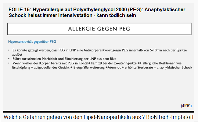
SLIDE 15: Hyperallergy to polyethylene glycol 2000 (PEG): Anaphylactic shock due to PEG always ends up in the intensive care unit - can be fatal
(original German: FOLIE 15: Hyperallergie auf Polyethylenglycol 2000 (PEG): Anaphylaktischer Schock wegen PEG landet immer in der Intensivstation - kann tödlich sein)
So, the last point is, that is also a bit discussed in the media, the allergy to the polyethylenglycol PEG (48'54 ''). I already said that the PEG itself is not toxic to us, it is completely harmless for our cells (49'0 ''). But there is some kind of hypersensitivity to the PEG (49'3 '').
Slide: ALLERGY TO polyethylene glycol 2000 (PEG)That means, people who I have come into contact with PEG at some point in his or her life (49'10 '') - no idea where that can be found, this have to know toxicologists (49'17''), well it could be that this person has already developed an antibody response to PEG (49'24''), and when sometimes in life another confrontation comes with PEG (49'29''), then will be a hypersensitivity reaction within 5 to 10 minutes and this can be super dangerous (49'36''). Well there is - yes - ok, these antibodies being formed against PEG first will be active then (49'47''), the lipid nanoparticles who are camouflaging the PEG are attacked by the antibodies and first of all the lipid nanoparticles are eliminated from the blood instantly (49'59''), and with this, the vaccination is completely without any effect (50'2''). Well, people with this immune response - there will be no spike protein formed any more (50'8''). There cannot come any immune response any more (50'10''). Oh yes, this was all in vein (50'13''). But now there is a problem with this allergy response (50'18''), because this is a reaction, but this is an allergic reaction, and this goes always with exhaustion, swollen face, dilation of blood vessels, shortness of breath, and sometimes an increased death rate (50'31 ''), in short: anaphylactic shock (50'34 ''). And we just know that with some people - I believe it was in Alaska State in the "USA" where this vaccination was sopped the first time (50'40''), because many people had an allergic reaction against PEG (50'46''). Well, there are photos circulating of many people being exhausted (50'50'') and with a swollen face (50'51'').
Hypersensitivity to PEG
-- It could be shown that PEG in lipid nanoparticles (LNP) triggers an antibody response against PEG within 5-10 minutes after the injection
-- Leads to rapid morbidity and elimination of lipid nanoparticles LNP from the blood
-- If the body has already come into contact with PEG before, e.g. with the second injection => allergic reactions such as exhaustion + swollen face + dilation of blood vessels + shortness of breath + increased death rate = anaphylactic shock
FOLIE 15: Hyperallergie auf Polyethylenglycol 2000 (PEG) - anaphylaktischer Schock, kann tödlich sein
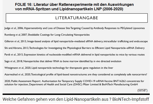
SLIDE 16: LITERATURE CREDITS
Judge et al., 2006, Hypersensitivity and Loss of Disease Site Targeting Caused by Antibody Responses to PEGylated Liposomes
Romberg et al, 2007: Sheddable Coatings for Long-Circulating Nanoparticles
Gilleron et al, 2013: Image-based analysis of lipid nanoparticle-mediated siRNA delivery, intracellular trafficking and endo.escape
Shi and Abrams, 2013: Technologies for Investigating the Physiological Barriers to Efficient Lipid Nanoparticle-siRNA Delivery
Partid et al., 2015: Expression kinetics of nucleoside-modified mRNA delivered in lipid nanoparticles to mice by various routes
Sago et al., 2018: Nanoparticles that deliver RNA to bone marrow identified by in vivo directed evolution
Witzigmann et al., 2020: Lipid nanoparticle technology for therapeutic gene regulation in the liver
Azarnezhad et al., 2020: Toxicological profile of lipid-based nanostructures: are they considered as completely safe nanocarriers?
2020: Public Assessment Report: Authorisation for Temporary Supply COVID-19 mRNA Vaccine BNT162b2 concentrate for solution for injection Department of Health and Social Care (DHSC) Pfize3r Limited & BionTech Manufacturing GmbH (50'55'')
Teilen / share:
Video from Dr. Vanessa Schmidt (December 25, 2020): What are the dangers of lipid nanoparticles? BioNTech vaccine (52'22 '') -
Video von Dr. Vanessa Schmidt (25.12.2020): Welche Gefahren gehen von den Lipid-Nanopartikeln aus ? BioNTech-Impfstoff (52'22'') --
SLIDE 1: RNA needs a lipid envelope, otherwise the RNA is "broken down" in 10 minutes - 4 lipids - The components of the Pfizer lethal injection: 4 lipids + substances for the K value + for the osmotic pressure -
SLIDE 2: The properties that lipid nanoparticles (LNP) must have -
SLIDE 3: The mRNA must be kept stable in the body: Lipid "ALC-0159" - The lipid: ALC-0159 = 2 - [(polyethylene glycol) -2000] -N, N-ditetradecylacetamide -
SLIDE 4a. Prerequisite: fusion and penetration into cell:
SLIDE 4b. Cholesterol on the mRNA vesicle
SLIDE 5: The mRNA is in the cell and must now be freed from the lipid envelope -
SLIDE 6a (drawing): Drawing of the spike protein in a body cell, pH values 7.2 and 7.4 -
SLIDE 6b (drawing): Drawing of the spike protein with reactions with APOE molecule and apolipo protein etc. -
SLIDE 6c (drawing): The spike protein is in the cell with a pH of 6.3 -
SLIDE 6d (drawing): The vesicle has pH 4.7 - is positively charged and the cap of the cholesterol is gone -
SLIDE 6e (drawing): The vesicle bursts with the inflow of water and the mRNA and lipids are free in the cell - SLIDE 6f (drawing): The burst vesicle in the cell: Toxic substances are circled: the cationic lipid and the mRNA - -
SLIDE 7a + b: Where do the lipid nanoparticles go? -
SLIDE 8: Rats injected 3 times in 2 weeks with gene "vaccination" from Pfizer - and the immune response - The immune response: Increased production of lymphocytes in the bone marrow - Migration to the spleen - Maturation and enlarged spleen + lymph nodes - The inflammatory reaction at the injection site : Instead of muscle cells there is only incrustation (connective tissue) - the muscle is restricted - the muscle cells: 13 different cell types - the lipid nanoparticles destroy cell walls - blood vessel ruptures occur - bleeding - and the fibrinogen wants to clot the exiting blood - the tissue is Destroyed after just 3 weeks - culprit lipid nanoparticles -
SLIDE 9: The structure of the liver - the copper cell in the liver vein - The damaged liver cells: vacuolization (the liver becomes holey like an Emmental cheese) -
SLIDE 10: The liver cells and the vacuolation (the liver becomes holey like an Emmental cheese) - Cholesterol is broken down in the liver - therefore the liver damage -
SLIDE 11a: Graphic of the spike protein with cholesterol, and a liver cell with receptors for the cholesterol - After the Pfizer gene vaccination: The lymphocyte level is low for 1 week - Lymphopenia - People who have been vaccinated should be quarantined for 1 week -
SLIDE 12: Massive decrease in lymphocytes in the first week - Lymphopenia in the intensive care unit -
SLIDE 13: 3 theses as to why the number of lymphocytes decreases: 1. LNP in the spleen - 2. LNP in the spinal cord - 3. B and T cells absorb LNP - Thesis 1: Concentration of lipid nanoparticles in the spleen - the spleen becomes damaged - Thesis 2: Concentration of the lipid nanoparticles in the bone marrow - Limitation of lymphocyte production in the bone marrow - Thesis 3: The lipid nanoparticles destroy the B and T cells - Formation of new B and T cells -
SLIDE 14: Reasons for cell death after Pfizer lethal injection: 1. Lipid nanoparticles produce oxygen radicals + cell chaos - 2. provoke DNA breaks in the lungs + spleen - 3. provoke thrombosis or hemolysis in the blood (red blood cells gone!) - 1. Production of Oxygen radicals Toxic effect: The lipid nanoparticles LNP provoke the production of oxygen radicals in the cell - Oxygen radicals in body cells change the calcium concentration - this changes the activity of proteins - Oxygen radicals in body cells can activate the cancer gene B53 - Oxygen radicals in body cells provoke oxidative stress - Oxygen radicals in the body cells provoke the release of cytokines - oxygen radicals in the body cells can break the DNA - too many DNA breaks provoke cell death - repair prevention - cancer cells - oxygen radicals in the body cells provoke interactions: the folding of the protein is missing or incorrect - protein becomes ineffective s - lipid peroxidation: cationic lipid components react with the cell - cell membrane becomes rigid - ions + water fill the cell until it bursts - cell death - 2. massive inflammatory reactions in the tissues / organs lipid nanoparticles cause dead tissue and inflammation - DNA breaks in Lungs, spleen, - Blood processes: Thrombosis and haemolysis (red blood cells have APOE receptors) - Haemolysis symptoms - Clinical studies with the Pfizer lethal injection: No tests for red blood cells or hormones -
EPISODE 15: Hyperallergy to polyethylene glycol 2000 (PEG): Anaphylactic shock due to PEG always ends up in the intensive care unit - can be fatal -
SLIDE 16: LITERATURE CREDITS -
Population watch: There is NO higher mortality 2020
Germany: https://countrymeters.info/de/Germany
Austria: https://countrymeters.info/de/Austria
Switzerlandz: https://countrymeters.info/de/Switzerland
Sources
[web01] IPT-Blutkrankheit: https://en.wikipedia.org/wiki/Immune_thrombocytopenic_purpura
[web02] https://de.wikipedia.org/wiki/Idiopathische_thrombozytopenische_Purpura
Photo sources


^
